- Pilot guides
- Supported guides
- Zygoma & Pterygoid guides

Surgical Guides

Home / Surgical Guides

One of the recent advancements in the field of digital implant dentistry is the use of surgical guides. This allows the dentist to place an implant in the most accurate and prosthetically favourable position. Thereby improving precision and reducing the patient’s chair time.

Our services

  • Pilot guide/Fully supported guide
  • Teeth supported guide/ Mucosa supported/ Bone supported
  • Zygoma & Pterygoid guides

How to approach us?

Mail the CBCT scan in DICOM format & dental cast scan in STL format via we transfer to ddscochin@gmail.com
or
Courier the data in a portable storage device (CD/Pendrive) to our address
or
Send the patient directly to the center along with the dental cast.

How do we respond?

We will dispatch the guide within 7 working days.

Work Flow

1

Starting Point

Send the patient directly to our center along with the dental cast. / Mail the CBCT scan in DICOM format & dental cast scan in STL format via we-transfer to ddscochin@gmail.com Or Courier the data in a portable storage device (CD/Pen-drive) to our address.
2

Assessment

Assessment of the data by our team for planning the implant.
3

Approval

.....interactions with the dentist regarding the virtual plan and approval
4

Fabrication

Once the plan is approved by the client we proceed with the fabrication of guide.
5

Despatch

Within 7 working(minimum) days the guide is dispatched to the client's address.

FAQ

  • Pilot guide/Fully supported guide
  • Teeth supported guide/ Mucosa supported/ Bone supported
  • Zygoma guides

Mail the CBCT scan in DICOM format & dental cast scan in STL format via we-transfer to ddscochin@gmail.com or courier CD to our center or send the patient directly to the center along with the dental cast.
Note: Make sure you are sending only the raw DICOM data and not the processed images with the viewer.

We will dispatch the guide within 7 working days.

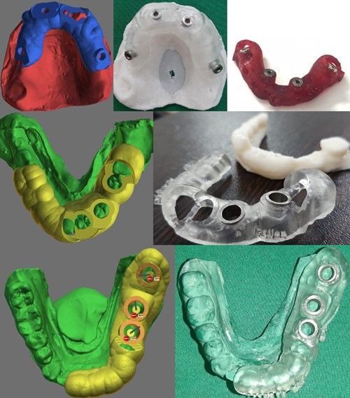
Edentulous CBCT dual scan protocol for guided implant planning

  1. Embed radiographic markers into the patient's denture.
  2. Prepare a putty bite index to hold the denture firmly in position and separate the teeth.
  3. Scan patient wearing the denture along with the putty index.
  4. Scan the denture alone, making sure that the dentures are in anatomical orientation.
  5. Please make sure that you are sending the CBCT data of the denture scan and not an STL file

(please contact the center if you require any further information about dual scan protocol)

Copyright © 2021-2023. Digital Dental Solutions, Kochi : ddskochi.com | A Dentaura Design